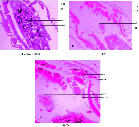
Figure 1. .
Cross sections of 4th instar larvae of Aedes aegypti treated or untreated with the Fusarium oxysporum extract. (A) Control was compared with (B) 100X and (C) 400X treated larval tissues, showing vacuolated gut epithelium (epi), gut lumen (lu), adipose tissue (ad) muscles (mu) nucleus (nu) and fat body (fb).
Notes: Larval mid-gut section was stained with Ehrlich’s haematoxylin, stained mid-gut tissues were viewed and photographed under light microscope at 100X and 400X magnification.

