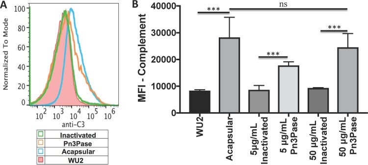
FIG 4.
Effect of Pn3Pase treatment on complement deposition on S. pneumoniae surface. Shown are the results from analysis of mouse complement deposition on Pn3Pase-treated or untreated type 3 S. pneumoniae by flow cytometry. The histogram (A) and quantification by mean fluorescent intensity (MFI) (B) of FITC-A were calculated from gating of Hoechst-positive cells. Statistical significance was determined with the two-tailed Student's t test. ***, P < 0.001; ns, not significant.

