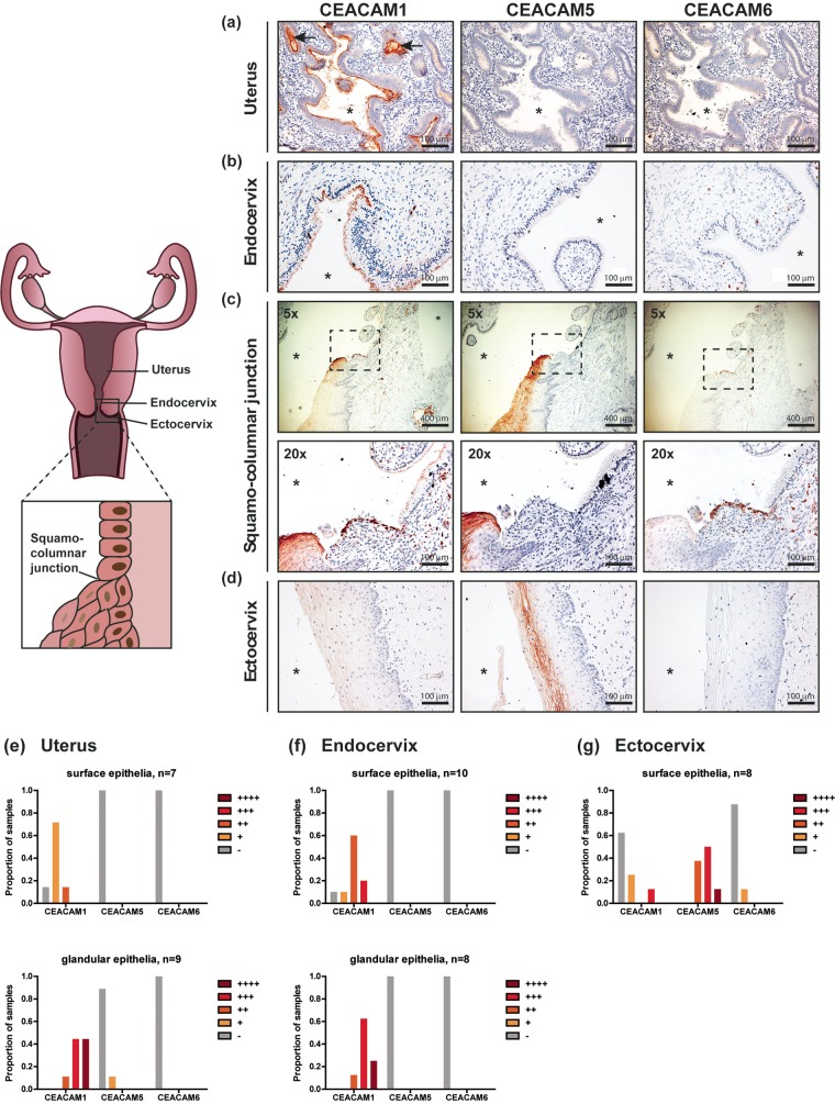
FIG 1.
Expression of CEACAMs along the human female reproductive tract. (a to d) Hysterectomy samples were stained with mouse monoclonal antibodies raised against human CEACAM1, CEACAM5, or CEACAM6 (red-brown), as indicated. Nuclei were counterstained with hematoxylin (purple). Images were obtained at a ×20 magnification unless otherwise specified. Asterisk, lumen; arrows, uterine glands. (e to g) The CEACAM staining intensity (red-brown) was qualitatively scored to distinguish low (+), moderate (++), strong (+++), and very strong (++++) levels of expression in individual patient samples. The proportion of individuals with the respective levels of each CEACAM is depicted graphically. −, undetectable; n, the number of individual patient samples that were examined for CEACAM expression on surface/glandular epithelia.

