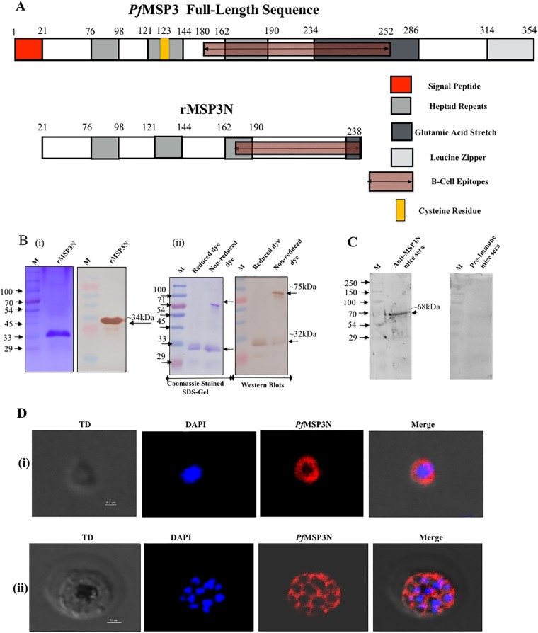
FIG 1.
Expression and localization of rMSP3N in P. falciparum asexual blood stages. (A) Schematic diagram showing the organization of PfMSP3. Numbers represent amino acid positions. (B) Coomassie-stained SDS-PAGE gel and immunoblot showing the purified rMSP3N are shown at left (i). At right, an SDS-PAGE gel and Western blot show migration of rMSP3N in reduced and nonreduced dye (ii). Lane M, molecular mass marker. (C) Western blot showing recognition of native PfMSP3 antigen in merozoite lysate by mouse anti-rMSP3N antibodies. (D) Confocal images showing immunostaining in asexual blood stage of P. falciparum using anti-rMSP3N mouse serum. Row i, merozoite stage; row ii, schizont stage. TD, transmitted light channel. Imaris software was used to convert confocal images into clear informative schematics.

