Fig. 3.

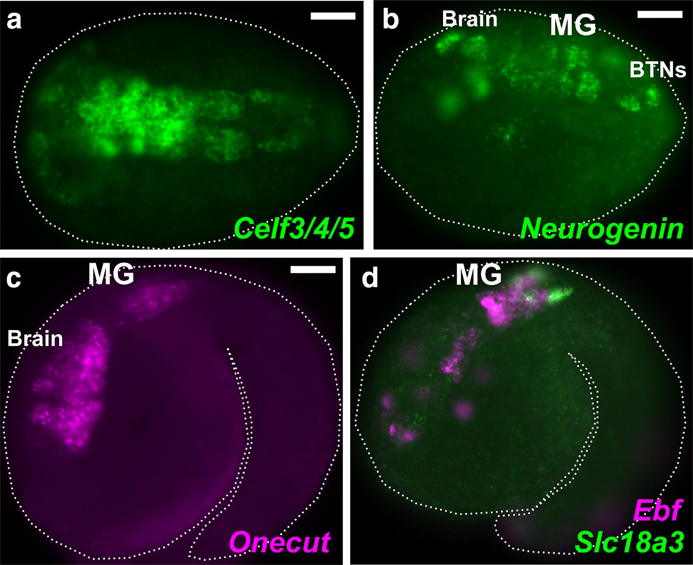
The developing central nervous system of Molgula occidentalis. a Dorsal view of a neurula stage Molgula occidentalis embryo. Green shows fluorescent in situ hybridization for Celf3/4/5 (also known as Etr-1) transcription. b In situ hybridization for Neurogenin in initial tailbud embryo, showing transcription in the embryonic brain, motor ganglion (MG), and bipolar tail neurons (BTNs). c In situ hybridization for Onecut, expressed in brain and MG. d Two-color fluorescent in situ hybridization showing co-expression of Ebf (magenta) and Slc18a3 (also known as VAChT, green) in the MG. Embryo outlined by dotted lines. Anterior is to the left in all images. All scale bars = 25 µm
