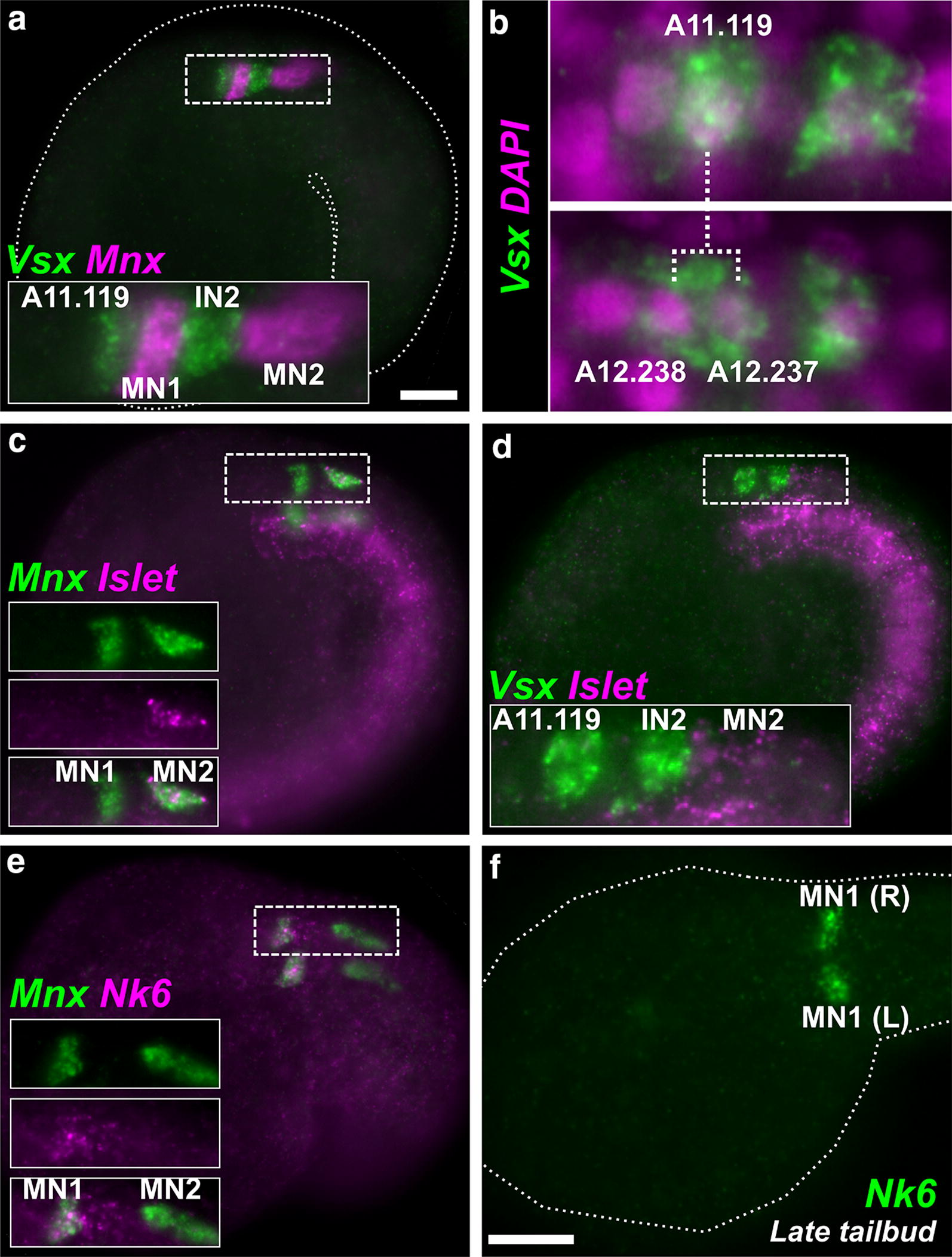
Fig. 4.

Motor neurons and interneurons in the MG. a Lateral view of mid-tailbud stage Molgula occidentalis embryo (dotted outline). Two-color fluorescent in situ hybridization reveals alternating expression of interneuron marker Vsx (green) in undifferentiated A11.119 interneuron progenitor cell and interneuon 2 (IN2) and motor neuron marker Mnx (magenta) in motor neurons 1 and 2 (MN1, MN2). Only one side of embryo is shown. Inset is a magnified view of dashed box. b In situ hybridization for Vsx (green) in embryos at two successive stages. Vsx is initially activated in A11.119 (top panel), which divides and gives rise to daughter cells A12.238 and A12.237, both of which continue to express Vsx at this stage. These data confirm that Vsx is activated in the A11.119 cell in M. occidentalis, which is not the case in Ciona (see text for details). Nuclei are counterstained with DAPI (magenta). c Two-color fluorescent in situ hybridization for Mnx (green) and MN2 marker Islet (magenta), revealing identities of MN1 (Mnx+/Islet-) and MN2 (Mnx+/Islet+) cells. Boxed area magnified in insets. d Two-color fluorescent in situ hybridization for Vsx (green) and Islet (magenta). Only one side of embryo is in focus. e Two-color fluorescent in situ hybridization for Mnx (green) and MN1 marker Nk6 (magenta), confirming identities of MN1 (Mnx+/Nk6+) and MN2 (Mnx+/Nk6-). f Nk6 in situ hybridization (green) in late tailbud embryo, showing sharpened expression in MN1 cells on both sides of the MG (R = right side and L = left side). All scale bars = 25 µm
