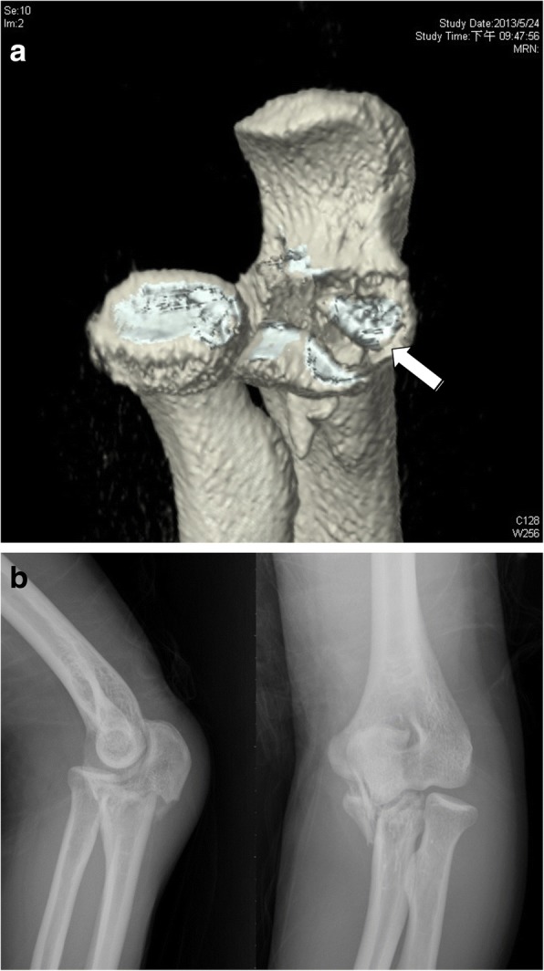
Fig. 2.

Two cases of anteromedial facet fractures. a Three-dimensional computed tomography scans showing an articular depression fracture (white arrow) close to the base of the coronoid process. b Plain radiographs showing an olecranon fracture with a displaced anteromedial facet fracture of the coronoid process
