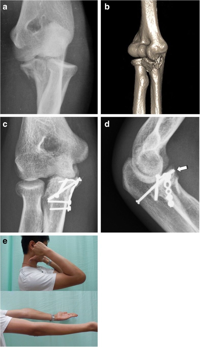
Fig. 4.

Anteromedial facet fracture with varus instability. a Plain radiograph. b Three-dimensional computed tomography image showing a large coronoid fragment. c Postoperative anteroposterior radiograph. d Postoperative lateral radiograph; the white arrow shows retrograde screw fixation of a coronoid base fracture. e Patient photo at 10 weeks after surgery showing good range of motion
