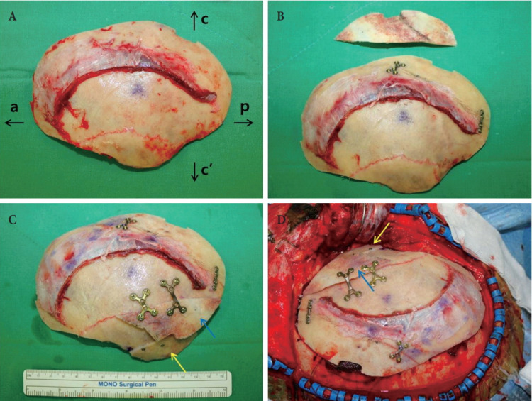
Fig. 2.

Surgical technique of temporal augmentation with calvarial onlay graft. A 48-year-old male with glioma on right temporal lobe underwent pterional craniotomy with temporal augmentation with calvarial onlay graft. (A)The cranial bone flap was separated. (B) Parietal segment of the cranial bone flap was marked and divided. The parietal segment was then split at the diploic space. The outer table was fixed it its original position. (C) The inner table was further divided and used as the flap for defect coverage (yellow arrow) and the onlay graft for temporal augmentation (blue arrow). (D) The reconstructed cranial bone flap was placed back on the patient. a, anterior; c, cephalic; p, posterior; c’, caudal.
