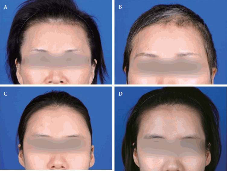
Fig. 3.

Preoperative and postoperative gross photographs. (A) Preoperative photograph of a 52-year-old female with meningioma on left frontal convexity who underwent pterional craniotomy with temporal augmentation. There was no temporal hollowing before surgery. (B) Postoperative photograph at 12 months of the patient without temporal hollowing. She was very satisfied with the results of the temporal augmentation. (C) Preoperative photograph of a 31-year-old female with meningioma who underwent pterional craniotomy without temporal augmentation. There was no temporal hollowing before surgery. (D) Temporal hollowing was seen at postoperative 12 months.
