
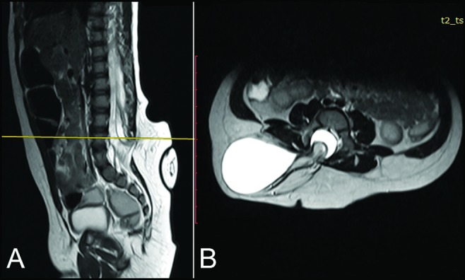
Figure 2.
Posterior fusion defects on L4, L5, and S1 levels and a 50 × 31 × 38 mm myelomeningocele sac, extending rightward from the defect: (A) Sagittal plane and (B) axial plan on level of L4 vertebrae
Official websites use .gov
A
.gov website belongs to an official
government organization in the United States.
Secure .gov websites use HTTPS
A lock (
) or https:// means you've safely
connected to the .gov website. Share sensitive
information only on official, secure websites.
Posterior fusion defects on L4, L5, and S1 levels and a 50 × 31 × 38 mm myelomeningocele sac, extending rightward from the defect: (A) Sagittal plane and (B) axial plan on level of L4 vertebrae