Figure 1.

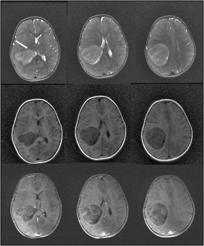
Preoperative MRI of the child showing the T2 images (top row), T1 images (middle row), and postcontrast T1 images (bottom row). The tumor abuts the corticospinal tract anteriorly (white arrow, top row)

Preoperative MRI of the child showing the T2 images (top row), T1 images (middle row), and postcontrast T1 images (bottom row). The tumor abuts the corticospinal tract anteriorly (white arrow, top row)