Fig. 2.
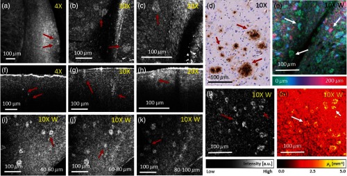
Amyloid-beta () plaques were investigated using visible light OCM at three different magnifications. (a)–(c) OCM en-face projection images taken with , , and magnification objectives, respectively in a 51-week-old mouse. (d) Immunohistochemically and hematoxylin stained tissue section of a plaque-rich region. Plaques appear brown. (e) Depth intensity projection over in a plaque-rich region in a 51-weeks-old mouse brain cortex. (f)–(h) Representative OCM B-scans taken with , , and magnification objectives, respectively. (i)–(k) OCM en-face projections over for different depth positions (40 to ) of a 64-weeks-old mouse brain. Plaques appear at different depths in the brain tissue. (l) OCM en-face projection image taken with the magnification water immersion objective of a 64-weeks-old mouse brain. (m) Attenuation image of (l). For all en-face projections, the intensity over the first underneath the tissue surface were averaged. In all images, representative plaques are marked with red and white arrows.

