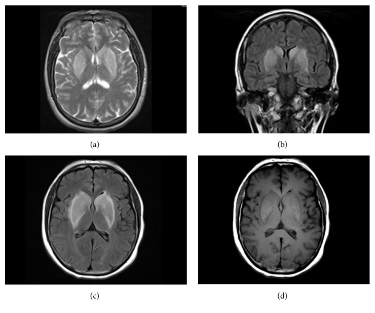
Figure 1.
Initial and follow-up MRI brain for Case 1. (a) Initial MRI: axial T2-weighted image with hyperintensities in the bilateral basal ganglia. (b) Initial MRI: coronal FLAIR-weighted image with hyperintensities in the bilateral basal ganglia. (c) Follow-up MRI: axial FLAIR-weighted image with hyperintensities in the bilateral basal ganglia. (d) Follow-up MRI: axial T1-weighted image with hyperintensities in the bilateral basal ganglia.

