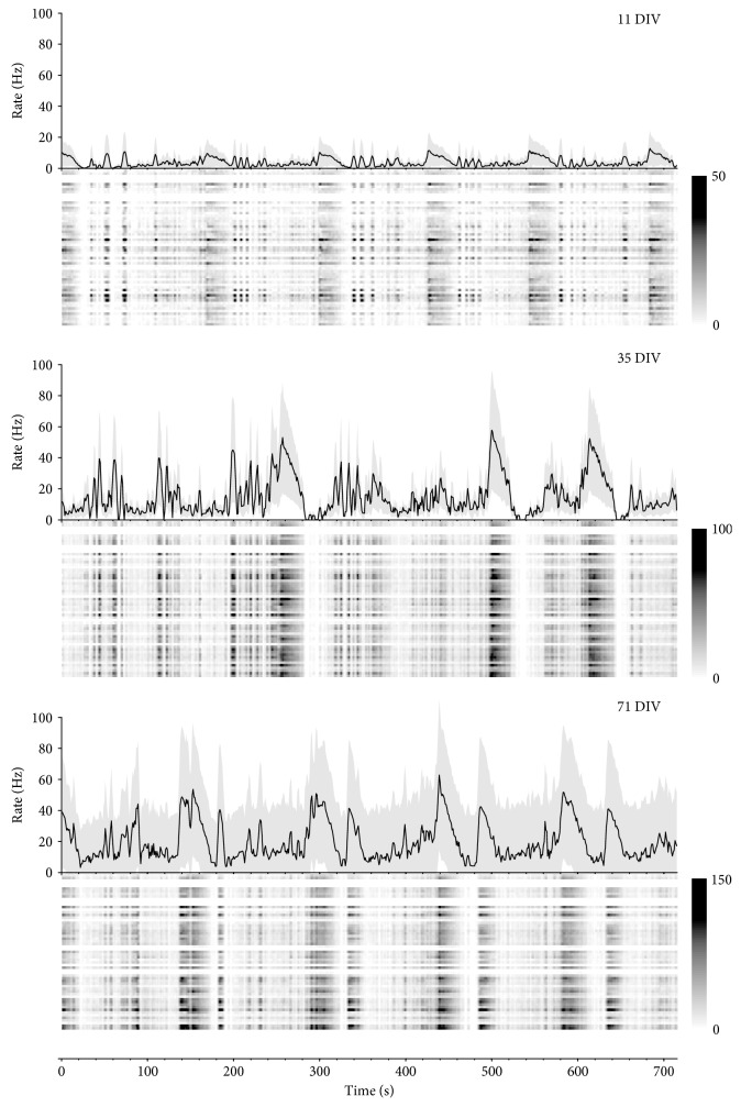
Figure 6.
The diversity of activity patterns of developing neuronal networks. In each subplot, the upper panel shows array-wide spike detection rates per second at different ages of the same culture (DIVs are labeled in the top right corner). The spike detection rates of individual electrodes in the array are shown with raster plots in grayscale (rows: electrodes). Color bars indicate spike rates (Hz). Shades in the line graphs show standard deviations of rates among channels of the corresponding array.

