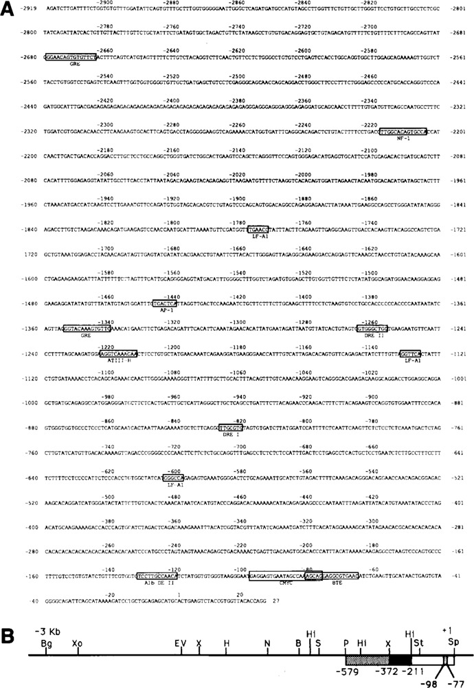
Figure 1.

Structure of the rat CYP2B2 promoter region. In A the sequence of 2919 bp of the promoter is shown. The location of putative protein factor binding sites is indicated, as well as the region showing homology with the LINE 1 element (discussed in the text). In B the physical map of the 5′-flanking region of the CYP2B2 gene is given, ranging from the transcription initiation point (+1) to 2919 bp upstream. B = BamH I, Bg = Bgl II, H = Hind III, Hi = Hinc II, N = Nco I, P = Pst I, S = Sma I, Sp = Sph I, St = Stu I, X = Xba I, EV = EcoR V, Xo = Xho I. Fragments used as probes in bandshift analyses are boxed. The location of the sequence from −98 to −77 of the proximal part of the CYP2B2 promoter showing homology with a c-myc regulatory element (see text) is indicated.
