
Figure 4.
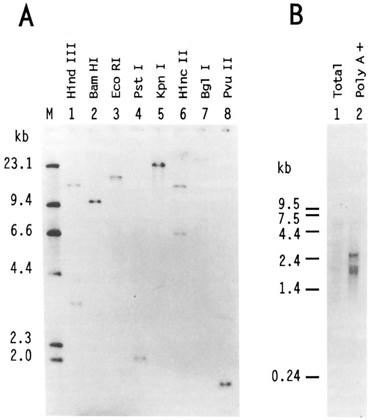
Southern and Northern analysis of human USF2. A. In each lane, 5 μg of HeLa DNA, digested by various restriction endonucleases as indicated, were resolved on a 0.8% agarose gel along with a 32P-labeled Hind III digest of λ DNA as size markers (lane M). After transfer to nitrocellulose, the blot was probed with the BamH I fragment corresponding to nucleotides 33 to 510 of hUSF2-C. Hybridization of this probe was observed with single genomic DNA fragments of 9.6 kb, 17 kb, 2 kb, 23 kb, 25 kb, and 1.7 kb after digestion with BamH I, EcoR I, Pst I, Kpn I, Bgl I, and Pvu II, respectively (lanes 2–5, and 7–8). Hybridization was also observed with 15.5- and 3.3-kb Hind III fragments (lane 1) and 13.6- and 6.5-kb Hinc II fragments (lane 6). The size of the DNA markers (in kb) is indicated at left. B. 2 μg of either total (lane 1) or poly(A)-selected HeLa RNA (lane 2) were separated on 1.2% agarose-formaldehyde gel and transferred to nylon membrane. The radioactive probe was the same as in A. The size (in kb) and migration of RNA markers run on the same gel is indicated at left.