
Figure 5.
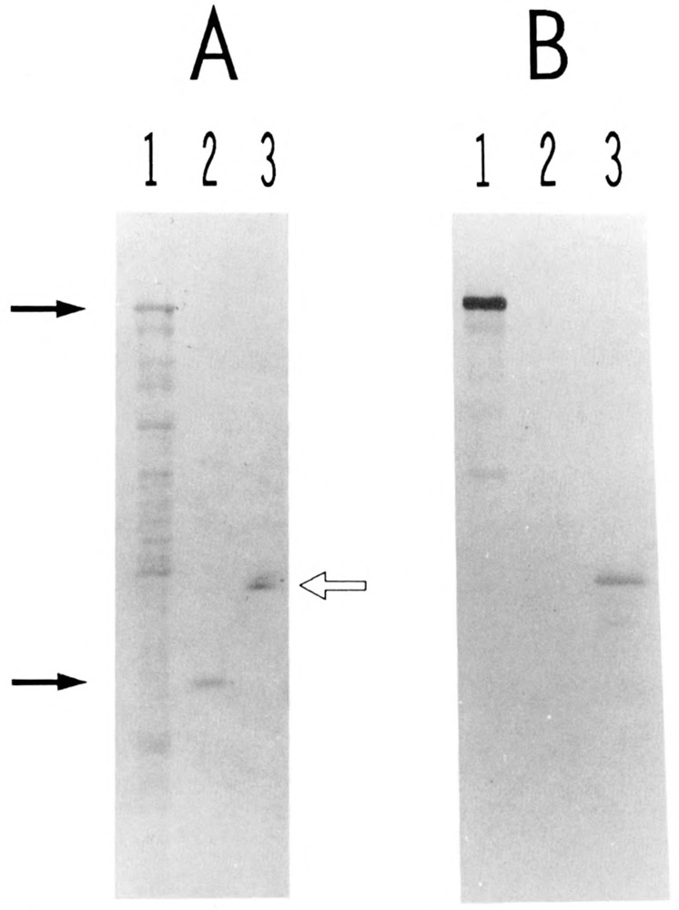
Presence of USF2 in purified preparation of HeLa USE The various protein samples were separated by SDS gel electrophoresis and transferred to nitrocellulose. One portion of the filter was stained with Fast Green (A). The rest of the filter was treated with immune rabbit serum to USF2, and specific binding of the antibodies was visualized with alkaline phosphatase-conjugated goat antibodies to rabbit IgGs (B). Lanes 1: proteins from 3 μl of an IPTG-induced culture of E. coli cells transformed with plasmid PUR-hUSF2 (see Materials and Methods). Lanes 2: 340 ng of purified His6-delUSF protein (see Materials and Methods). Lanes 3: 160 ng of purified HeLa USF (Mono S fraction). The two arrows at left indicate the respective migration of the β-galactosidase-USF2 and polyhistidine-USF1 fusion proteins. The open arrow at right indicates the migration of the 44- and 43-kDa polypeptides characteristic of purified HeLa USF preparations (Sawadogo et al., 1988).