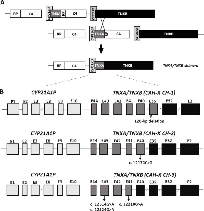
Fig. 2.
Schematic diagram of the TNXA/TNXB chimeric genes. Most common is a bimodular RP-C4-CYP21-TNX region. TNXB encodes the active tenascin-X gene (black) and TNXA encodes the corresponding pseudogene (dark grey). CYP21A2 encodes the active 21-hydroxylase gene (medium grey) and CYP21A1P encodes the corresponding pseudogene (light grey). Panel A shows formation of a TNXA/TNXB chimeric gene due to misalignment during meiosis resulting in deletion of the CYP21A2 gene. Panel B is a schematic of exons (rectangles) of representative TNXA/TNXB chimera. TNXA/TNXB chimeric genes have been classifed into 3 types (CH-1 to CH-3) based on the junction site location. The 120-bp deletion at the boundary of exon 35 and intron 35 of TNXB is shown by an open triangle and present in CAH-X CH-1. The c.12174C>G pseudogene variant in Exon 40 identifies CAHX CH-2, and CAH-X CH-3 is characterized by a cluster of 3 pseudogene variants: c.12218G>A in exon 41, and c.12514 G>A, and c.12524 G>A in exon 43. Adapted from reference [78]; with permission.

