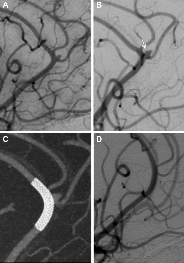
FIGURE 1.

A, Right ICA angiography shows a small right pericallosal artery aneurysm with the neck involving the origin of the right callosomarginal artery. Patient underwent several angiograms for follow-up of this aneurysm and a previously treated large unruptured basilar tip aneurysm with stable size of the pericallosal aneurysm (not shown). B, Five-year follow-up angiogram showed aneurysm growth with new pseudolobulation in its posterior aspect (arrow). C, Immediate postintervention cone-beam CT with multiplanar 3-D reconstruction shows adequate aneurysm neck coverage and slight straightening of the pericallosal artery. D, Twelve-month follow-up angiography shows complete aneurysm occlusion with patency of covered branches.
