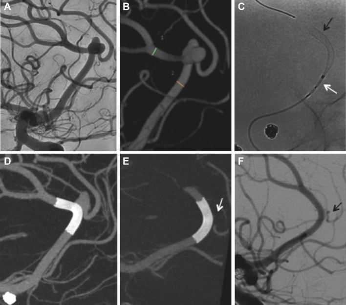
FIGURE 2.
A and B, Right ICA angiography and 3-D rotational angiography reconstruction reveal an anterior cerebral artery azygos configuration with an unruptured bilobed aneurysm located at the pericallosal and callosomarginal bifurcation. C, A 2.5 × 16 mm PED is successfully deployed across the neck of the pericallosal/azygos right anterior cerebral artery (black arrow at distal aspect of PED and white arrow at proximal portion still within delivery system). D, Cone-beam CT with multiplanar 3-D reconstructions confirms proper vessel wall apposition of the PED. E, Six-month follow-up cone-beam CT with multiplanar 3-D reconstruction shows complete occlusion of the bilobed aneurysm (arrow pointing to expected location of aneurysm). F, Corresponding angiography confirms patency of the parent vessel with narrowing of covered branch, which remains patent (arrow). Note the change in configuration with slight straightening of the pericallosal artery at 6-mo follow-up (F) when compared to initial study (A).

