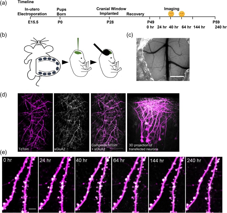
Figure 1.
Repeated in vivo imaging of doubly transfected layer 2/3 neurons of M1 cortex. (a) Experimental time course. Morning sessions are marked with a symbol. (b) Embryos from E15.5 timed pregnant C57Bl6 fmr1 HET mice were injected with a mixture of pUB-SEP-GluA2-WPRE and pCAG-tdTom DNA constructs into the lateral ventricles and neurons were transfected using an electrode tweezer. (c) A cranial glass window implanted over the motor cortex. Scale bar: 1 mm. (d) 2PLSM in vivo images of transfected region of cortex showing overlap of tdTom (magenta) and sGluA2 (white) along with 3D projection of a Z of the same region. Scale bar: 100 μm. (e) Repeated in vivo imaging of apical dendrites of layer 2/3 neurons in M1 cortex showing stable expression of tdTom and sGluA2 over the experimental duration. Scale bar: 5 μm.

