Fig. 1.

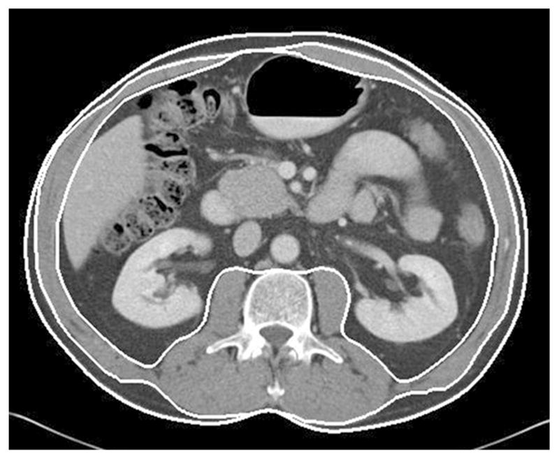
Axial CT image of the abdomen at the third lumbar level. Boundary lines between subcutaneous fat, skeletal muscle, and visceral fat compartments are shown (lines are thickened for ease of visualization)

Axial CT image of the abdomen at the third lumbar level. Boundary lines between subcutaneous fat, skeletal muscle, and visceral fat compartments are shown (lines are thickened for ease of visualization)