
Figure 2. Analysis of VLA-4 and E-selectin ligand expression on mo-DCs.
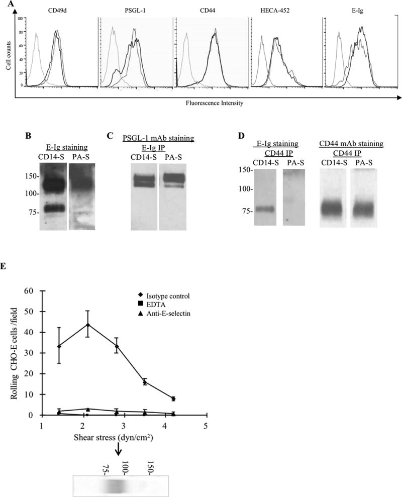
(A) Representative flow cytometry analysis of mo-DCs staining using mAbs to CD49d (VLA-4), PSGL-1 and CD44, and HECA-452 mAb and E-Ig. Grey lines represent isotype control (or, for E-Ig, staining in the absence of Ca2+), dotted black line represents PA-S mo-DCs and solid black lines are CD14-S mo-DCs. (B–D) Analysis of glycoprotein E-selectin ligands expressed by mo-DCs: (B) Equivalent amounts of cell lysates of CD14-S and PA-S mo-DCs were resolved by SDS-PAGE electrophoresis, and immunoblotted with E-Ig chimera. Two E-selectin-reactive bands were visible in lysates of CD14-S mo-DCs (~130 kDa and ~80 kDa), whereas only one band (~130 kDa) was reactive on PA-S mo-DCs. (C) Equivalent amounts of cell lysates of CD14-S and PA-S mo-DCs were immunoprecipitated with E-Ig, and immunoprecipitates were then electrophoresed and immunoblotted with anti-PSGL-1 mAb. (D) CD44 immunoprecipitates (CD44 IP) from equivalent amounts of cell lysates of CD14-S and PA-S mo-DCs were immunoblotted with E-Ig chimera or anti-CD44 mAb. (E) Blot rolling assay of CD44 immunoprecipated from CD14-S mo-DCs. CD44 immunoprecipitates were resolved by SDS-PAGE, blotted and stained with anti-CD44 mAb (blot below the graph). E-selectin-transfected CHO cells (CHO-E) were perfused over blots at 1.4 dyn/cm2 and then shear stress was increased to 4.2 dynes/cm2. E-selectin-dependent tethering and rolling was observed at the CD44 band (arrow). Assays were performed in the presence or absence of 5 mM EDTA, or following preincubation of CHO-E with isotype control mAb or function blocking anti-human E-selectin mAb (clone 68-5H11).