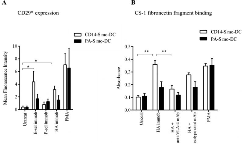
Figure 3. VLA-4 binding activity is triggered by engagement of CD44 on CD14-S mo-DCs.

(A) Flow cytometry analysis of activated β1-integrin expression. Expression of the activation-dependent epitope of CD29 (HUTS-21) was assessed on CD14-S mo-DCs (white bars) and PA-S mo-DCs (black bars) incubated on plates coated with either E-selectin, P-selectin, or HA, or stimulated with PMA. Graph values represent the mean fluorescence intensity of HUTS-21 mAb staining (CD29*) of mo-DCs (n= 3, mean ± SD). (B) Adhesion of CD14-S and PA-S mo-DCs to CS-1 fibronectin fragment. Mo-DCs were incubated on uncoated plates or plates coated with HA, and then collected for analysis of binding to CS-1 peptide coated on plates. Integrin activation by treatment with PMA was used as positive control; the numbers of CS-1-adherent cells were quantified by light absorbance (595 nm) following crystal violet staining. As shown in the Figure, binding to HA by CD14-S mo-DCs, but not by PA-S mo-DCs, induced VLA-4 adhesion to CS-1 peptide, which was abrogated by treatment with anti-VLA-4 blocking mAb (HP2/1). Values are means ± SD (n= 3). Statistically significant differences (P ≤ 0.05) related to HA engagement are indicated by brackets and asterisks.
