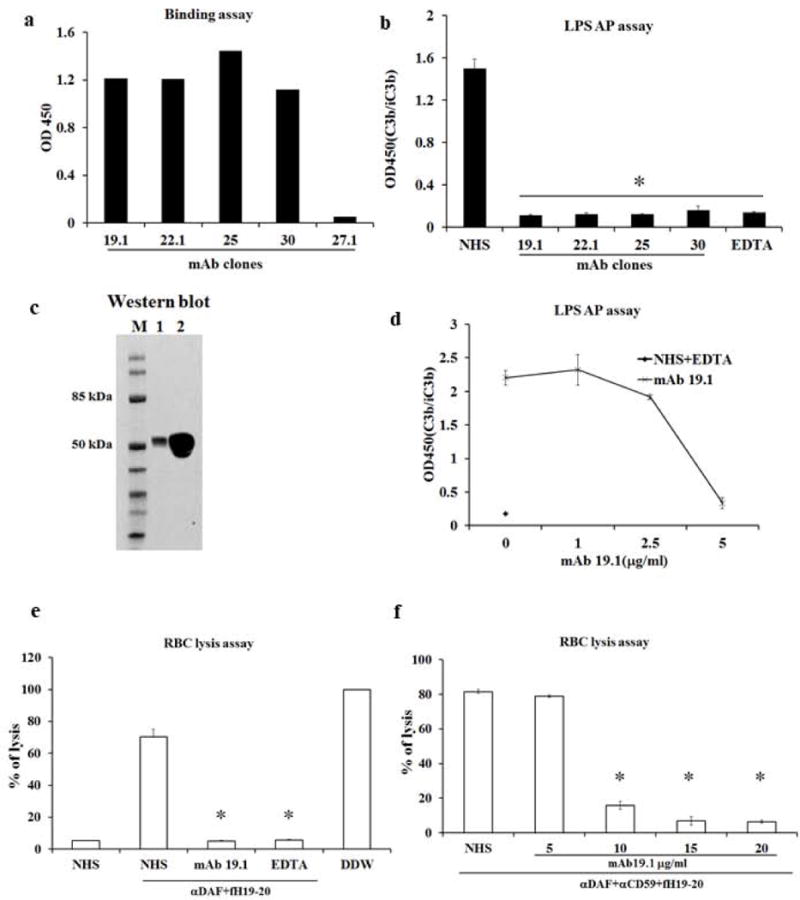
Figure 1. Generation and characterization of anti-human properdin antibodies.

a. Screening of anti-human P mAbs by ELISA. Culture supernatant from 4 hybridoma clones, mAb 19.1, 22.1, 25 and 30, showed strong binding to plate-immobilized hP, where supernatant from a negative mAb clone 27.1 showed no binding. b. Inhibition of LPS-induced AP complement activation by affinity purified mAb 19.1, 22.1, 25 and 30. All 4 clones of mAbs effectively inhibited AP complement activation when added to 10% normal human serum (NHS) at a final concentration of 1μg/ml. A sample with EDTA added (EDTA) served as a positive control for inhibition. A sample with no mAb added (NHS) served as the baseline AP complement activation. OD450 value represents ELISA reading of relative C3 fragments deposition (i.e. activated and plate-bound C3). c. Western blot confirming that mAb clone 19.1 specifically recognizes purified properdin and properdin in NHS. Lane 1: 20 ng of purified human properdin; Lane 2: 1μl of NHS separated on non-reducing SDS-PAGE. mAb 19.1 was used at 2 μg/ml in Western blotting. d. Further functional characterization of mAb clone 19.1 showing it concentration-dependently inhibiting LPS-induced AP complement activity in 50% NHS. NHS was diluted 1:1 (i.e. to 50%) in EGTA-Mg+2 supplemented GVB++ buffer and then pretreated with or without mAb (1, 2.5 and 5μg/ml) for 1hr at 4°C before adding to LPS-coated plates. It completely blocked AP complement activity at 5μg/ml in this assay. EDTA serum (NHS+EDTA) was used as a positive control for complement inhibition. e, f. Human erythrocytes were lysed by 50% NHS in the presence of human factor H SCR19-20 and anti-DAF antibody (7.5 μg/ml, panel e) or anti-DAF and anti-CD59 antibodies (10 μg/ml for both, panel f). Lysis was prevented by EDTA or mAb 19.1 (5 μg/ml for panel e and as indicated in panel f). Assays were performed in triplicates and percent lysis was normalized to hypotonic lysis (100%) with distilled water (DDW). All data are representative of at least three independent experiments and values are expressed as mean (SD) of at least 3 replicate assays per data point. * p<0.001 compared with sample without mAb 19.1 treatment (NHS). One-way ANOVA.
