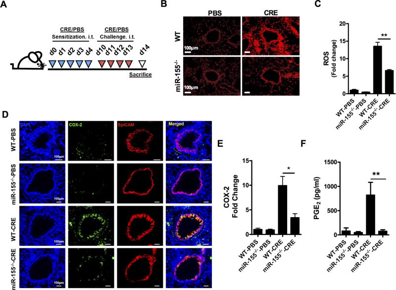
FIGURE 5.
miR-155 regulates epithelial COX2 expression levels in a mouse model of asthma. (A)Experimental model of cockroach allergen-induced asthma. (B) Representative images of dihydroethidium-stained (DHE-stained) airways of PBS- or CRE-challenged WT and miR-155−/− mice. Scale bar: 100 µm. (C) Quantitative data for ROS expression. (D) Co-localization of COX-2 (green) and EpCAM (red) in the lung sections of WT and miR-155−/− mice by immunofluorescence staining. Nuclei were stained with DAPI (blue). Scale bar: 100 µm. (E) Quantitative data for COX-2 expression. (F) Levels of PGE2 in BALFs of PBS- or CRE-treated WT and miR-155−/− mice. Quantification of gene expression in lung tissues was performed with ImageJ v1.50e. n=9–12 mice/group pooled from two independent experiments. Data represent the mean ± SEM. *P<0.05, **P<0.01.

