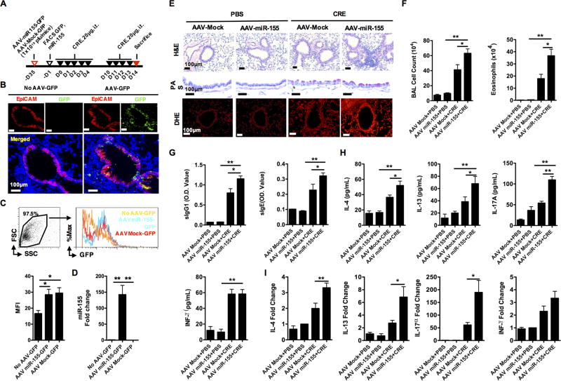
FIGURE 7.
miR-155−/− mice infected with AAV-miR-155 reversed the decreased lung inflammation in cockroach allergen-induced mouse model. (A) Experimental setup for mice infected with AAV-miR-155-GFP or AAV-Mock-GFP and subsequent mouse model of asthma. (B) Detection of GFP (green) in airway epithelial cells (red) of AAV-GFP infected or non-infected mice by immunostaining. Nuclei were stained with 6’-diamidino-2-phenylindole (DAPI, blue). Scale bar: 100 µm. (C) Detection of AAV-GFP in lung tissue cells of AAV-GFP infected or non-infected mice by flow cytometry. (D) RT-PCR analysis of miR-155 expression in lung tissues of AAV-infected or non-infected mice. (E) Paraffin tissue sections of lung tissues from PBS- or CRE-challenged AAV-Mock or AAV-miR-155 infected miR-155−/− mice were stained with H&E (upper panel), Periodic Acid-Schiff (PAS, middle panel), and DHE (lower panel). Scale bar: 100 µm. (F) Mouse BALF total and eosinophil cell counts. (G) Serum levels of cockroach allergen-specific IgE and IgG1. (H) Levels of cytokines in BALFs. (I) RT-PCR analysis of Th1, Th2, and Th17 cytokines in the lung tissues. n=6–10 mice/group pooled from two independent experiments. Data are expressed as the mean ± SEM. *P<0.05, **P<0.01.

