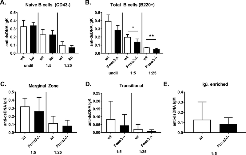
Figure 6: Foxo3−/− B cells do not secrete increased autoantibodies.

A-D) B cell subpopulations were isolated from wild type and Foxo3−/− mice on the FVB background and stimulated with 5 ug/ml LPS for 5 days. anti-dsDNA Igκ and Igλ antibodies were measured by ELISA in the indicated dilutions of culture supernatant. Results are shown for Igκ as mean +/− SD, n = 4–6. *p < 0.05, **p < 0.01. Igλ anti-DNA antibodies were not detected. Subpopulations analyzed are as follows: (A) naïve B cells purified by negative selection with anti-CD43 beads; (B) total B cells purified by positive selection with anti-B220 beads; (C) marginal zone (B220+CD21hiCD23-/lo) and (D) transitional (B220+CD21-CD23-) B cells isolated as described in Materials and Methods. (E) Igκ-depleted B cells (and thus Igλ+) were isolated from spleens of wild type and Foxo3−/− mice on the FVB background and stimulated with 5 ug/ml LPS for 5 days. Anti-dsDNA IgM (E) and IgG (not detected and thus not shown) antibodies were measured by ELISA in the indicated dilutions of culture supernatants. Results are presented as mean +/− SD, n = 7.
