
Figure 1. Effect of Pyk2-deletion and estrogen on OB cell number and proliferation.
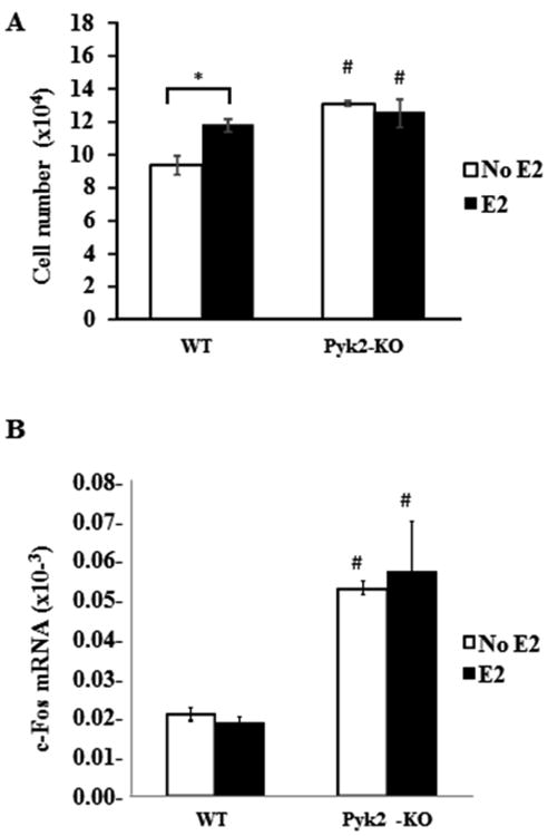
A) WT and Pyk2-KO OBs from neonatal calvaria were plated at 5×10⁴ cells and cultured in the presence or absence of 100 nM E2. Cells were counted after 4 days. B) QPCR analysis was used to determine c-fos mRNA expression in WT and Pyk2-KO OBs cultured for 4 days in the presence or absence of 100 nM E2. 18S was used as the housekeeping control to normalize the mRNA transcript under investigation. The 18S Ct values were WT (14.26 ± 0.09) and Pyk2-KO (14.56 ± 0.21). The data are shown as mean of ΔCT ± SEM of triplicate or quadruplicate samples. Statistical significance of p<0.05 is indicated (*) for effects within a genotype and (#) for significant changes for the same treatment between WT and Pyk2-KO OBs.