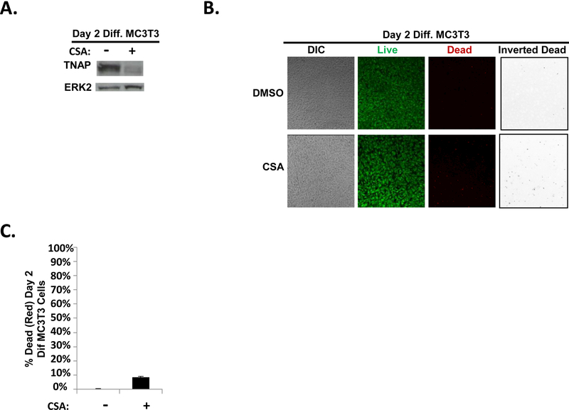
FIGURE 3. CSA pretreatment of early differentiating MC3T3 osteoblasts reduces TNAP expression.
(A) MC3T3 cells were treated as in Fig. 2A except whole cell lysates were harvested on Day 2 (without the addition of KG1a-CXCR4 AML cells, AMD3100, or SDF-1). Immunoblot depicting the effect of CSA on TNAP expression in MC3T3 cells. The same membrane was stripped and re-probed for total ERK2 as a control, n=3. (B) MC3T3 cells were treated as in Fig. 2a except live/dead staining and imaging occurred on Day 2 (without the addition of KG1a-CXCR4 AML cells, AMD3100, or SDF-1). Live/dead staining and confocal imaging of live (green) and dead (red) cells were used to ensure MC3T3 cell viability in the presence of CSA. Images were acquired on three separate days for a total of 15 images analyzed for each condition. (C) Statistical summary of (B). Bars depict mean results + S.E.M., n=3.

