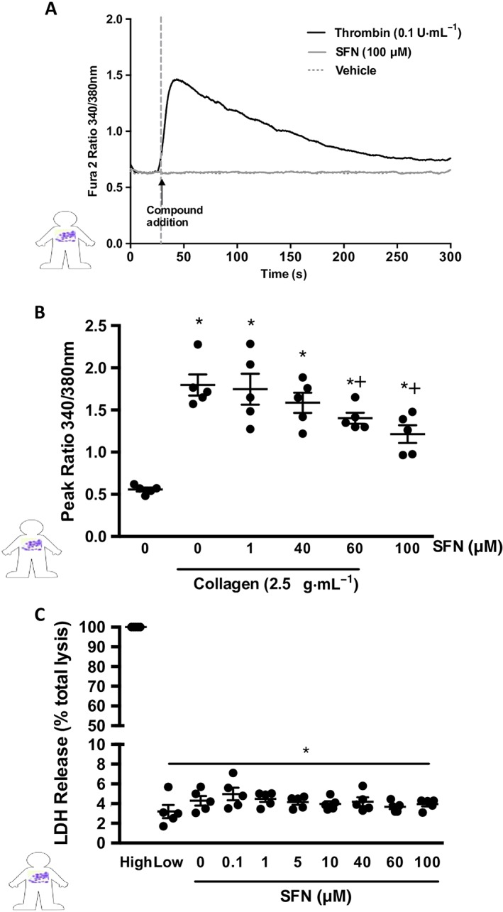
Figure 5.

SFN influence over collagen‐induced Ca2+ signalling. Cytosolic calcium release was measured using Fura 2AM dye in human platelets. (A) Representative trace showing temporal increase in Fura 2 AM fluorescence following administration of the positive control (0.1 U·mL−1 thrombin) but with no effect of SFN treatments alone. (B) Histogram of peak 340/380 nm emission, in platelets stimulated with 2.5 μg·mL−1 collagen following SFN treatment. (C) LDL release as a marker for cytotoxicity following SFN treatment, data presented as a % LDH release when compared to completely lysed cells. Data are mean ± SEM of n = 5 independent donors per group. * P < 0.05 versus un‐stimulated control. + P < 0.05 versus collagen stimulated vehicle group.
