Figure 5.
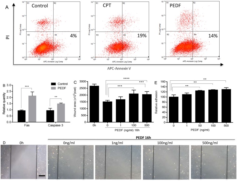
Dose response effect of PEDF on apoptosis, migration, and adhesion of mouse dermal endothelial cells (MDMEC). (A) Representative flow cytometric analysis of MDMEC exposed to PEDF or CPT. MDMEC were treated with PEDF (100 ng/ml) or CPT (5uM/positive control) for 24 hours. Early apoptotic cells were detected by flow cytometry using an annexin V apoptosis detection Kit. (B) Fas and caspase 3 mRNA expression after PEDF treatment. n = 3 for control Fas & caspase 3, and PEDF caspase 3; n = 6 for PEDF Fas. (C) In vitro migration of MDMEC following scratch wound placement with/without PEDF treatment. Wound area was quantified in pixels using ImageJ. Mean ± SD; n = 5 for 0 h, n = 5, 12, 12, and 15 for PEDF 0, 1, 100, and 500 ng/ml, respectively. (D) Representative images of cell migration in scratch wounds. Scale bar = 100 µm. (E) Adhesion of MDMEC with and without PEDF, assessed by measuring the quantity of remaining adherent cells following a 4 h exposure to PEDF. Mean ± SD; n = 3. **p < 0.01, ***p < 0.001, and ****p < 0.0001, Student’s t-tests was used for statistical analysis.

