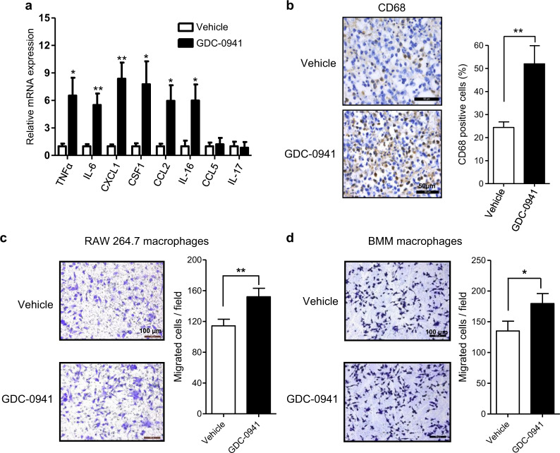
Fig. 2. GDC-0941 treatment enhanced macrophage infiltration and provoked pro-inflammatory molecules in 4T1 allografts.
a Quantitative RT-PCR analysis of a panel of cytokines and chemokines in 4T1 tumors of Balb/c mice following treatment with GDC-0941 (n = 5) or vehicle control (n = 5) for 3 days. Gene expression was normalized to β-actin. Error bars represent mean ± S.E.M. b Representative images of immunohistochemical staining for CD68 of 4T1 allografts after treatment with GDC-0941 or vehicle (n = 5 per treatment group) for 10 days. Scale bar, 50 μm. Percentages of positive cells of CD68 are shown. Error bars represent mean ± S.E.M. c, d Migration assay of RAW264.7 macrophages (c) and BMM macrophages (d) stimulated by conditioned media from 4T1 cells treated with 1 µM GDC-0941 or vehicle. Scale bars, 100 μm. Quantifications of migrated cells from three independent experiments are shown as mean ± S.D. *P < 0.05; **P < 0.01; ***P < 0.001 (Student’s t-test)

