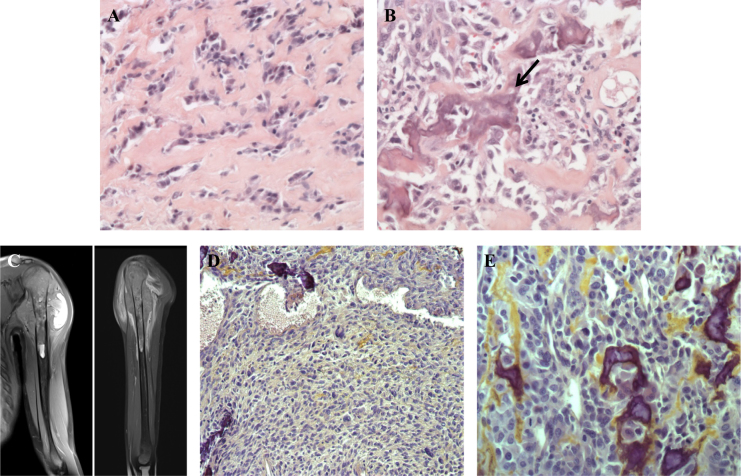
Fig. 1.
Hematoxylin and eosin staining of the first osteosarcoma showed predominant sclerosing areas with tumoral cords and lobules embedded in a network of dense collagen strands a. Few areas were made of more cellular proliferation intermingled with calcified osteoid (arrow, b). MRI of the left arm performed 10 years later showed a second osteosarcoma of the upper part of the humerus infiltrating the nearby soft tissue c. Compared with the first osteosarcoma, this second tumor harbored very different histological features that correspond to a conventional high-grade osteosarcoma, without sclerosing areas, with more frequent atypical nuclei and cells, and with a higher cellular density d, e

