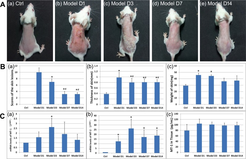
Figure 1.
DNFB-induced AD-like lesions in BALB/c mice and MT dynamics. (A) Clinical picture of DNFB-induced AD-like lesions. (B) Evaluation of DNFB-induced AD-like lesions. (a) Scores of the lesions. Skin lesions of erythema, edema, exfoliation, and scaling were scored as 0 (none), 1 (mild), 2 (moderate), and 3 (severe)17,18. *P < 0.01 versus model group on Day 1. #P < 0.01 versus model group on Day 3. (b) The thickness of the skin. Skin thickness was measured three times with three different sites of the back. The average values were presented. *P < 0.01 versus control group. #P < 0.01 versus model group on Day 1. (c) The weight of the skin. Dorsal skin measuring 6 mm × 6 mm at a fixed position across all the mice was sheared without subcutaneous fat. *P < 0.001 versus control group. (C) (a,b) The expression of the mRNA level of MT-1 and MT-2 in the skin tissue. The primary value of Ct was transferred into 2−ΔΔCt. *P < 0.01 versus control group. (c) The expression of the protein level of MT-1 in skin tissue.

