Figure 2.
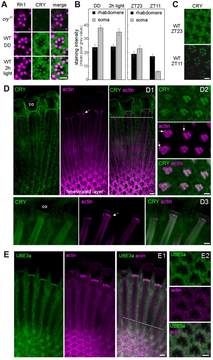
CRY is stably expressed in the rhabdomeres of the photoreceptor cells, co-localizes with actin but only marginally with Ubiquitin Ligase3. (A) Cross sections of one ommatidium, respectively, stained with anti-Rh1 (magenta) and anti-CRY (green). No CRY staining is present in cry01 mutants, whereas in wild-type (WT) flies CRY is detected in all eight photoreceptor cells including their rhabdomeres. After 2 h illumination with 1000 lux, rhabdomeric CRY staining did not disappear. (B) Quantification of CRY staining intensity in the rhabdomeres and photoreceptor somata of WT flies raised in constant darkness (DD), after subsequent 2-h exposure to 1000 lux and under a regular 12:12 light-dark cycles at the end of the night (ZT23) and end of the day (ZT11), respectively. Means (± SEM) of 12 independent retinas, respectively, are shown. In the rhabdomeres, CRY-staining was not reduced after 2-h light-exposure (p = 0.404) and only slightly at ZT11 during the regular light-dark cycle in comparison to the 2-h light exposure (p = 0.001). During the light-dark cycle, CRY staining of the rhabdomeres was the same at ZT23 and ZT11 (p = 1.0), but CRY staining in the somata of the photoreceptor cells was much lower at ZT11 than at ZT23 (p < 0.001). CRY staining in the somata of the photoreceptor cells was very high after keeping the flies in DD and was not significantly reduced after the 2-h light exposure (p = 0.275). (C) Examples of retinal CRY staining at ZT23 and ZT11. During the day (ZT11) CRY was significantly lower than during the night (ZT23) in the photoreceptor somata (p > 0.0001) but not in the rhabdomeres (p = 1.0). Statistics: one-way ANOVA followed by Tukey’s multiple comparisons test. (D) Co-localization of CRY and actin (visualized by fluorochrome-conjugated Phalloidin) in the retina in two longitudinal views (D1,D3) and one cross section (D2). The position of the cross section is indicated by the broken white line in D1. Actin-Phalloidin staining was variable, but always very strong in the fenestrated layer at the bottom of the retina (D1) and in the distal retina just below the cristal cones (co; arrows in D1–D3). In the latter place, actin surrounded the photoreceptor cells (arrows in D2). To a weaker extent, actin was also always present in the rhabdomeres, where it co-localized with CRY (D2, D3 and weakly in D1). (E) Co-localization of ubiquitin ligase 3a (UBE3a) and actin in the retina in a longitudinal (E1) view and a cross section (E2) at ZT23. The position of the cross section is indicated by the broken white line in (E1). UBE3a was highly expressed in the soma of the photoreceptor cells but only marginally in the rhabdomeres. Scale bars: 5 μm.

