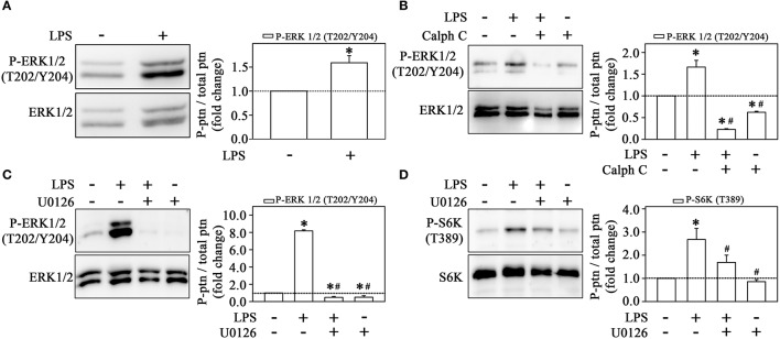
Figure 7.
Activation of the mTORC1/S6K pathway induced by LPS is mediated by ERK activation. LPS induces ERK phosphorylation in THP-1 cells (n = 3) (A). The effect of 10−6 M calphostin C (B) or U0126 (C) on LPS-induced ERK phosphorylation (n = 3). U0126 prevented the effect of LPS on S6K phosphorylation (n = 3) (D) The levels of phosphorylation were determined by the relationship between the optical density of specific phosphorylated residues and total fractions (left panels). The result is expressed in fold change in relation to an untreated control (bar graph). S6K, S6 protein kinase. Images are representative of three independent experiments. The results are presented as means ± SE. *p < 0.05 vs. unstimulated non-adhered cells (control). #p < 0.05 vs. LPS.

