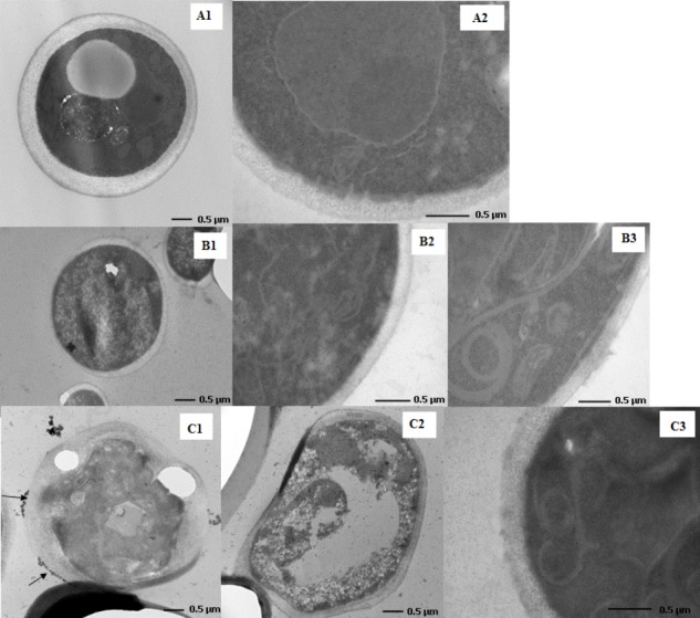
FIGURE 3.

Changes in the cell morphology of compound 1- and compound 2-treated Candida albicans. Transmission electron microscopy images of C. albicans ATCC 10231 strain in the absence (A1,A2) and presence [CoCl2(dap)2]Cl (B1–B3) and [CoCl2(en)2]Cl (C1–C3) complexes in MIC values.
