Figure 4.
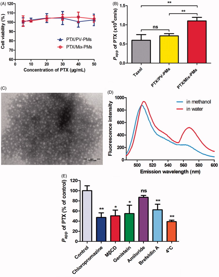
(A) Cytotoxicity of PTX/PV-PMs and PTX/Mix-PMs to Caco-2 cells after incubation for 3 h (n = 6). (B) Apparent permeability coefficients of PTX for different formulations (Taxol®, PTX/PV-PMs, and PTX/Mix-PMs) at 20 μg/mL PTX across Caco-2 cell monolayers from apical (AP) to basolateral (BL) side at 37 °C (n = 3). **p < .01, ns p > .05. (C) Transmission electron microscope images of basolateral medium collected after 3 h of incubation of Caco-2 cell monolayers with PTX/Mix-PMs. (D) Fluorescence emission spectra of basolateral medium collected after 3 h of incubation of Caco-2 cell monolayers with FRET micelles at excitation wavelength of 488 nm. (E) Transport of PTX across Caco-2 cell monolayers after 3 h of incubation with PTX/Mix-PMs under different conditions (n = 3). P app of PTX (% of control) indicated the percentage of P app to the control in the absence of any inhibitor at 37 °C. ns p > .05, *p < .05, and **p < .01 compared with the control.

