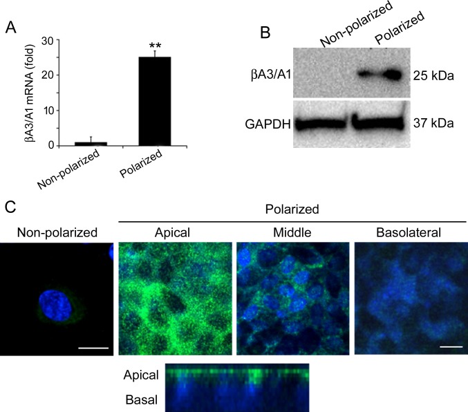
Figure 1.
βA3/A1-crystallin is differentially expressed in nonpolarized and polarized human RPE cells. (A, B) Polarization of RPE cells results in a highly significant increase in the mRNA and protein expression of βA3/A1-crystallin, compared to nonpolarized RPE cells. All values are represented as mean ± S.D. **P < 0.01 (Mann-Whitney U test), n = 4. (C) The distribution of βA3/A1-crystallin in polarized cultured human RPE cells as seen by confocal microscopy (upper) indicates that βA3/A1-crystallin is predominantly localized to the apical region of RPE cells. This was confirmed by a confocal cross-section image (lower). Scale bar: 20 μm for nonpolarized cells, 60 μm for polarized cells.

