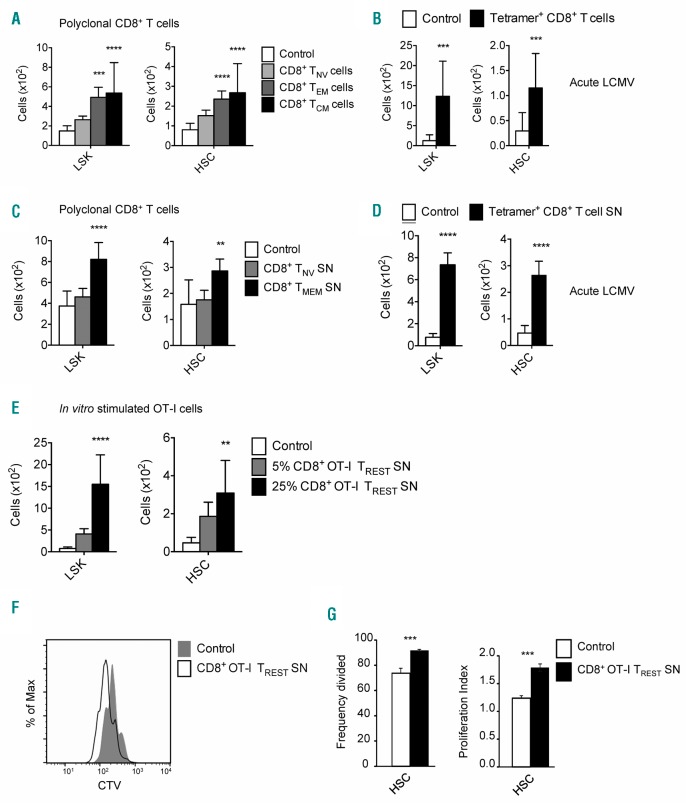
Figure 2.
Co-culture of HSCs with memory T cells or their supernatant propagates the expansion of hematopoietic stem cells (HSCs). (A) Absolute cell numbers of Lin−/loSca-1+cKit+ (LSKs) and HSCs generated in the assay when HSCs are co-cultured with the different subsets of the bone marrow (BM) CD8+ T-cell population. (B) Absolute cell numbers of LSKs and HSCs generated in the assay when HSCs are co-cultured with acute BM lymphocytic choriomeningitis virus (LCMV)-specific memory CD8+ T cells isolated based on recognition of GP33–41, GP276–286 and NP396–404 epitopes on day 34 post infection. (C) Absolute cell numbers of LSKs and HSCs generated in the assay when HSCs are cultured with supernatant (SN) derived from BM naïve (CD44−) and memory (CD44+) CD8+ T cells. (D) Absolute cell numbers of LSKs and HSCs generated in the assay when HSCs are cultured with SN derived from acute BM LCMV-specific memory CD8+ T cells isolated based on recognition of GP33–41, GP276–286 and NP396–404 epitopes on day 37 post infection. (E) Absolute cell numbers of LSKs and HSCs generated in the assay when HSCs are cultured with 25% or 5% SN derived from in vitro generated resting antigen-experienced CD8+ OT-I T cells. (F) Flow cytometry analysis of the dilution of Cell Trace Violet (CTV) dye by HSCs when cultured with control medium or in vitro generated resting antigen-experienced CD8+ OT-I T cells. (G) Frequency divided and proliferation index of HSCs that are cultured with SN derived from in vitro generated resting antigen-experienced CD8+ OT-I T cells. Graphs show mean±Standard Deviation of each tested condition (n=3–16 wells), representative of 2–4 independent experiments.

