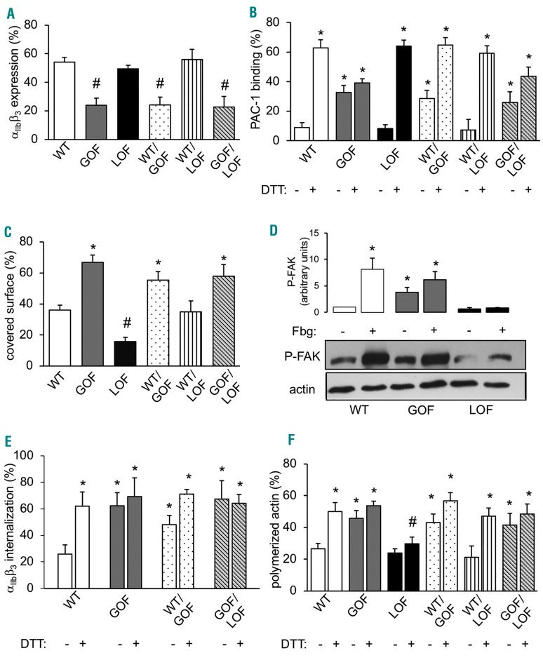
Figure 2.
αIIbβ3 expression and function in CHO cells. (A) αIIbβ3 expression measured by flow cytometry in CHO cells expressing wild-type (WT) β3 and/or gain-of-function (GOF) or loss-of-function (LOF) β3 variants in different combinations (#significantly decreased vs. WT; n=8; P<0.05). (B) PAC-1 binding measured by flow-cytometry in CHO cells expressing WT β3 and/or gain-of-function (GOF) or loss-of-function (LOF) β3 variants in different combinations. Full αIIbβ3 activation was obtained by incubation with DTT (25 mM) (*significantly increased vs. WT resting; n=8; P<0.01). (C) Spreading of WT and mutant β3-expressing CHO cells after 15 minutes of adhesion to fibrinogen-coated plastic coverslips (*significantly increased vs. WT; #significantly decreased vs. WT; n=5; P<0.05). (D) Focal Adhesion Kinase (FAK) phosphorylation of WT and mutant integrin β3-expressing CHO cells in suspension (-) and after adhesion to fibrinogen (+). Densitometric analysis was performed using the Image J software (*significantly increased vs. WT-; n=5; P<0.05). (E) Internalized αIIbβ3 in WT CHO cells and mutant CHO cells expressing wild-type αIIbβ3 and N331S β3, alone or in combination with WT or R786W β3, treated or not with dithiothreitol (DTT) (*significantly increased vs. WT resting; n=3; P<0.05). (F) Polymerized actin (F-actin) content of WT and of different mutant β3-expressing CHO cells measured by flow cytometry before and after stimulation with DTT (25 mM) (*significantly increased vs. WT; #significantly decreased vs. WT; n=5; P<0.05).

