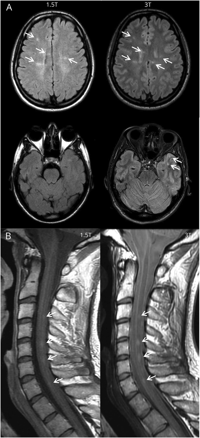
Figure 2. Lesion detection on 1.5T and 3T.

(A) Baseline fluid-attenuated inversion recovery brain imaging of a 28-year-old woman with clinically isolated syndrome presenting with optic neuritis. At 3T, more lesions were identified compared to 1.5T: periventricular lesions 6 vs 6, juxtacortical lesions 6 vs 3, and deep white matter lesions 12 vs 5. On both field strengths, no enhancing lesions were seen. This led to fulfilment of the 2017 revisions of the McDonald criteria for dissemination in space and not for dissemination in time at both 1.5T and 3T. Therefore the diagnosis of MS could not be made on both field strengths. So, even though high-field MRI increased the lesion detection of supratentorial demyelinating lesions in this patient, this did not change the diagnosis. (B) Baseline proton density spinal cord imaging of a 23-year-old man with clinically isolated syndrome, presenting with brainstem symptoms. Besides lesions in the brain, spinal cord lesions were identified on both field strengths. Even though there is a difference in image quality, an equal number of lesions were detected at 1.5T and 3T.
