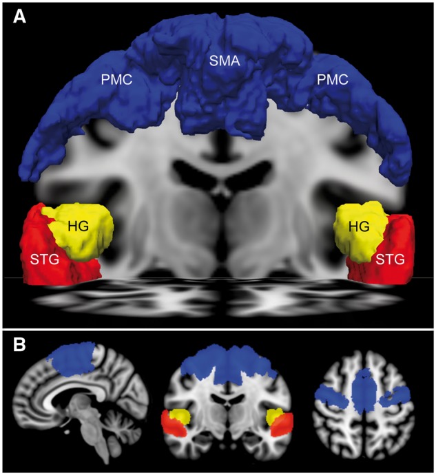
Figure 1.

Regions of interest for functional MRI analysis. (A) Anterior view of the supplementary motor area (SMA) and premotor cortex (PMC) region of interest (blue) used to assess motor imagery fMRI responses, as well as the Heschl’s gyrus (HG, yellow) and superior temporal gyrus (STG, red) regions of interest used to assess language and music functional MRI responses. All regions of interest are rendered in MNI152 space and superimposed upon a coronal image at the level of the mid-thalamus and an axial image at the level of the STG. (B) Sagittal (left), coronal (middle), and axial (right) images of the supplementary motor areas/premotor cortices, Heschl’s gyrus, and superior temporal gyrus regions of interest.
