
Figure 5.
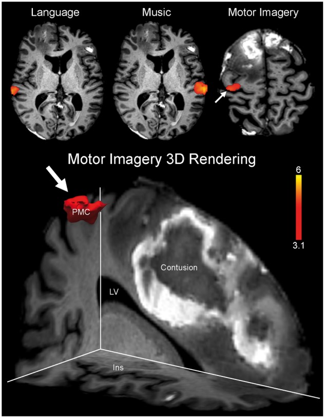
Functional MRI evidence of command-following in CMD. Functional MRI data are shown as Z-statistic images to demonstrate stimulus-specific responses in a patient whose behavioural evaluation suggested a vegetative state (Patient P14). Z-statistic images are thresholded at cluster-corrected Z scores of 3.1 (inset colour bar) and superimposed on T1-weighted axial images. There is functional MRI evidence of command-following on the motor imagery task (arrow), indicating CMD. In the bottom panel, a 3D rendering of the functional MRI response to the motor imagery task is shown (arrow). This response is located within the prespecified supplementary motor area/premotor cortex region of interest. Specifically, the response is located within the premotor cortex (PMC) in close neuroanatomic proximity to a right frontal contusion. The images in the top row are shown in radiological convention. Notably, despite functional MRI activation within the superior temporal gyrus during the language stimulus (top left), the patient was classified as having an absent response to language because of the absence of a response within Heschl’s gyrus. Ins = insula; LV = lateral ventricle.