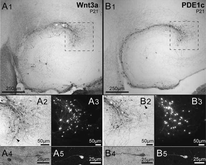
Figure 1.
Two lines of tdTomato reporter mice identify Cajal–Retzius cells. (A1) Low-magnification micrograph of immunolabeled cells of the Wnt3a mouse hippocampus. Notice the presence of cells along the hippocampal fissure and dentate gyrus outer molecular layer. As previously reported (Quattrocolo and Maccaferri 2014) notice also the presence of a few stained granule cells and hilar neurons. The dotted square identifies the CA3 pole region, which has the highest density of Cajal–Retzius cells (Anstötz et al. 2016). (A2,A3) Cajal–Retzius cells of the pole region shown at higher magnification in a DAB stained slice (dotted region of A1) and in a different preparation with fluorescence microscopy. Individual cells are shown in more detail in the (A4,A5) panels. Notice the typical “tadpole” morphology identifying the neurons as a Cajal–Retzius cells. (B1−B5) Sections obtained from PDE1c mice. Notice the clear labeling of Cajal–Retzius cells. Same organization as in (A1−A5).

