Figure 2.
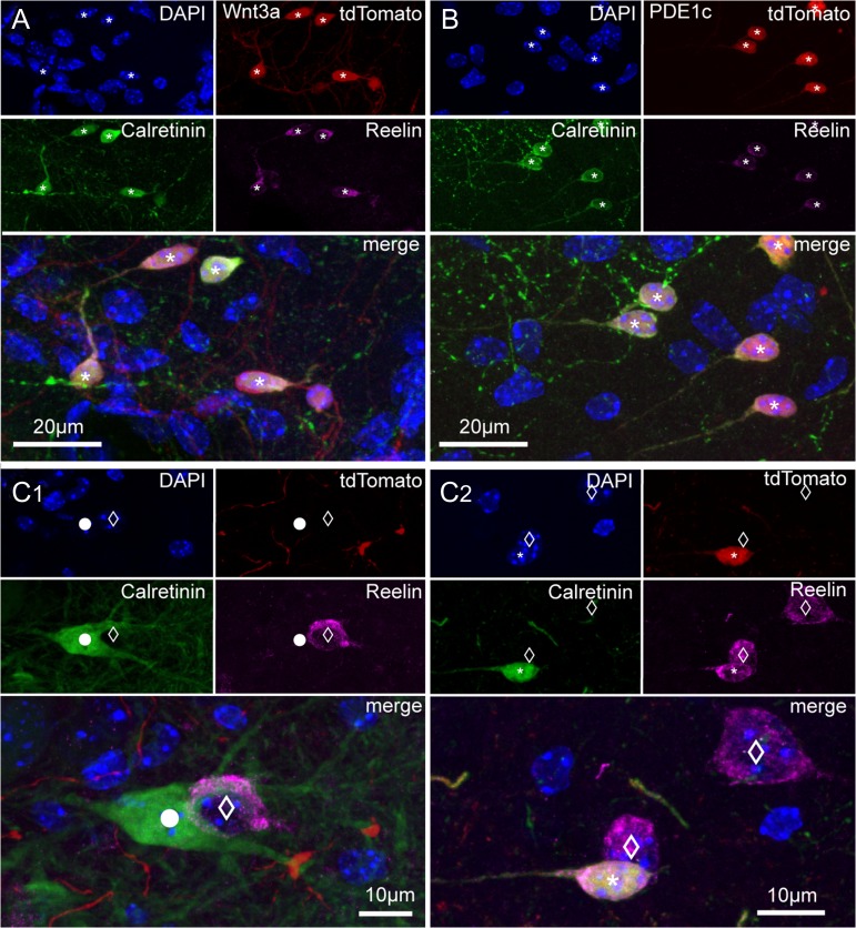
Immunoreactivity for calretinin and reelin in tdTomato-positive and negative cells of Wnt3a and PDE1c animals. (A) Images of the hippocampal fissure region from a juvenile Wnt3a mouse (P36) showing DAPI nuclear counterstaining and the labeling of tdTomato, calretinin, and reelin in single pictures, and then superimposed. Cells identified by the asterisks express all markers. (B) As in (A), but for the PDE1c mouse line (P36). (C1,C2) Calretinin and reelin immunoreactivity in cells that do not express tdTomato. (C1) Calretinin labeling of a putative hilar mossy cells (circle) and reelin positivity of a putative hilar GABAergic interneuron (empty diamond). DAPI nuclear counterstaining, tdTomato, calretinin, reelin labeling shown first as isolated images, and then superimposed. (C2) Reelin immunoreactivity in cells non expressing tdTomato in the hippocampal fissure region. DAPI nuclear counterstaining, tdTomato, calretinin, and reelin labeling of cells of the hippocampal molecular layers are first shown as single pictures and then superimposed. Notice the nontadpole-like morphology of the 2 reelin-positive cells (empty diamonds) that are not labeled by tdTomato and the tadpole-like appearance of the tdTomato, reelin, and calretinin-expressing cell (asterisk).

