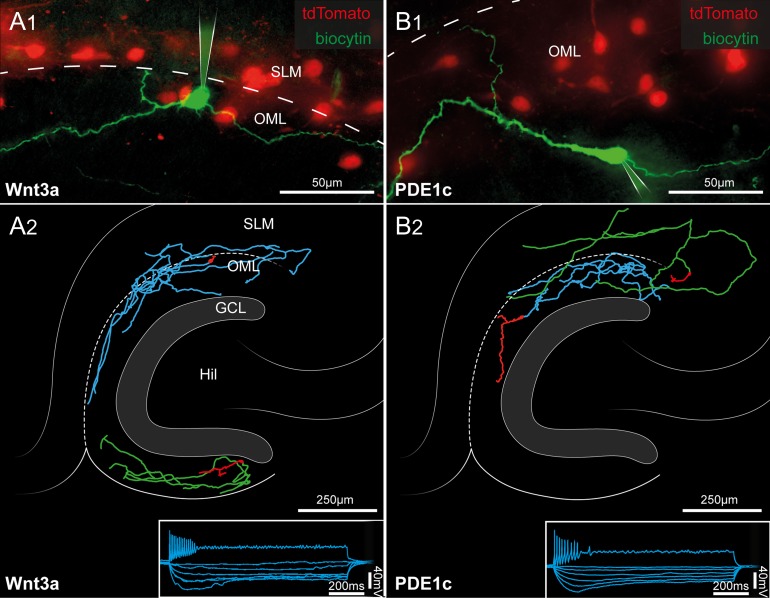
Figure 3.
Morphofunctional identification of tdTomato-expressing cells of the hippocampal molecular layers as Cajal–Retzius cells. (A1) Pseudo-color image showing a biocytin-filled Cajal–Retzius cell (green) in a slice obtained from a P15 Wnt3a mouse. Notice the typical emergence of the axon (arrowhead) opposite the main dendritic trunk. Remaining tdTomato cells are shown in red. SLM, stratum lacunosum-moleculare; OML, outer molecular layer of the dentate gyrus. The dotted line marks the hippocampal fissure. (A2) Post hoc anatomical reconstructions of 2 recorded Cajal–Retzius cells form Wnt3a mice. Notice the typical axonal arborization in the hippocampal molecular layers and the lack of a complex dendritic tree. Hil, hilus; GCL, granule cell layer. The bottom right inset shows the electrophysiological response of a labeled cell to a series of current steps (1 s duration, −25, −20, −15, −10, −5, and 65 pA). (B1,B2) Same experiments and analysis as in (A1,A2), performed on slices from P13 PDE1c mice.

