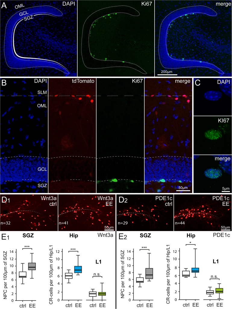
Figure 4.
Parallel impact of enriched housing conditions on hippocampal neurogenesis and Cajal–Retzius cell density. (A) Identification of proliferating cells in the subgranular zone (SGZ) of the dentate gyrus by Ki-67 immunoreactivity. Left panel: DAPI counterstaining, middle panel: Ki-67-labeled nuclei, and right panel: previous images superimposed. OML, outer molecular layer; GCL, granule cell layer. (B) Simultaneous identification of dentate gyrus structure (left: DAPI counterstaining), Cajal–Retzius cells (middle left: tdTomato fluorescence), dividing cells (middle right: Ki67), and superimposition of all the images (right: merge). SLM, stratum lacunosum-moleculare. (C) High resolution image of a Ki-67-labeled nucleus in the SGZ. Top, middle, and bottom panels show DAPI counterstaining, Ki-67 immunoreactivity, and the 2 images overlapped, respectively. (D1) Comparison of Cajal–Retzius cell densities in a control Wnt3a mouse (ctrl) versus an animal housed in environmental enriched conditions (EE). Notice the increased numbers of Cajal–Retzius cells in the image from the treated mouse (n = 32 vs. 41, respectively). (D2) Same as in (D1), but for PDE1c animals. (E1) Summary plots showing the densities of Ki-67-positive nuclei of neural progenitor cells (NPC) (left panel) and of Cajal–Retzius cells (in the hippocampus: Hip and in layer 1 of the adjacent cortex: L1, right panel) of Wnt3a mice housed in control (ctrl) and environmental enriched (EE) conditions (left panel). (E2) Same analysis as in (E1) carried on in sections from PDE1c mice.

